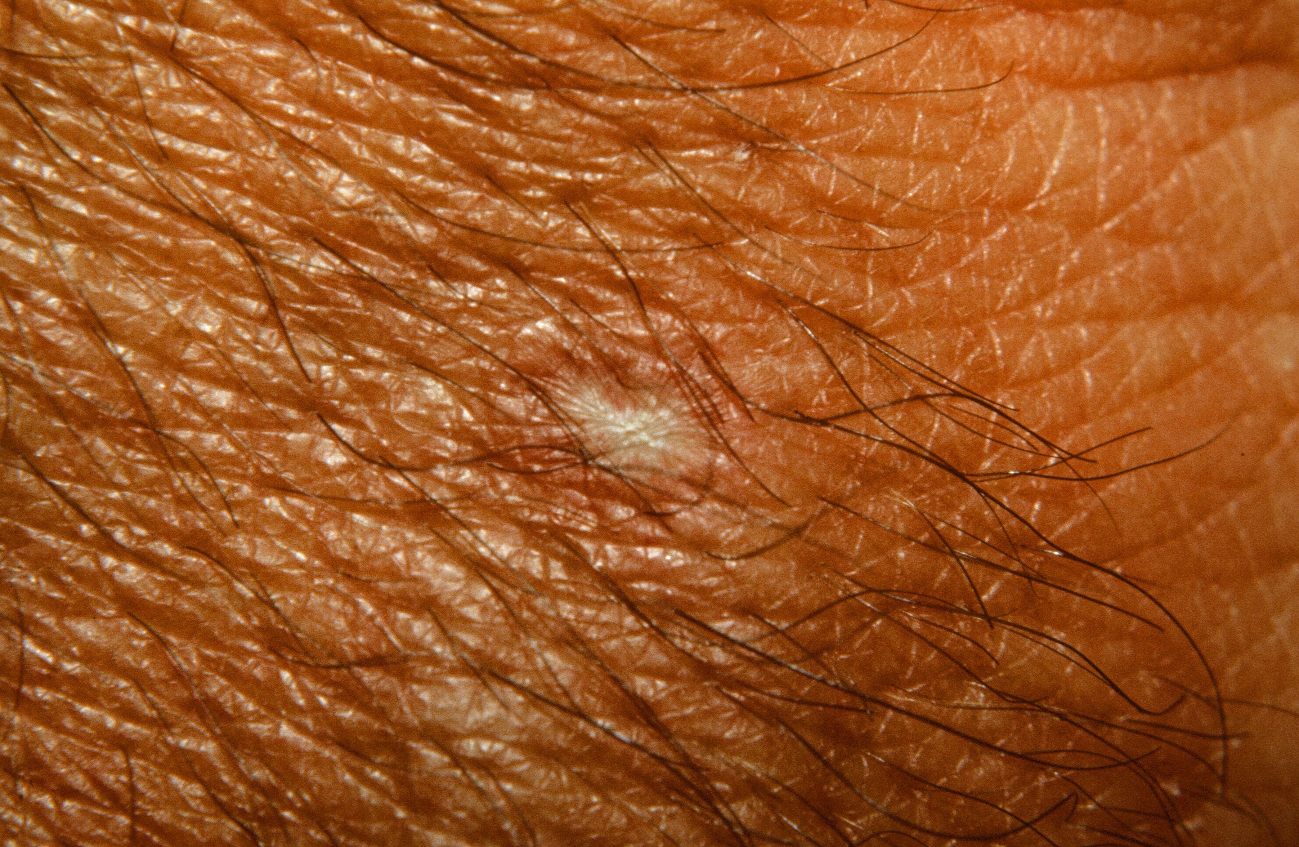
An atrophic, white, porcelain-centered lesion on the wrist in Dego's Disease
An atrophic, white, porcelain-centered lesion on the wrist in Dego's Disease
Also known as Dego's disease (DD), atrophic papulosis (AP) is an occlusive vasculopathy that presents as porcelain white areas. There is a benign cutaneous form and a malignant systemic (and usually fatal) form. Some patients have been found to have abnormalities of the fibrinolytic system or of their platelets. Positive antiphospholipid antibodies have been found. Multiple members of one family were reported affected in a pattern suggestive of autosomal dominant inheritance.
Erythematous papules on the trunk and proximal extremities which develop into atrophic, white, porcelain-centered lesions in a young- to middle-aged man are characteristic. A wedge-shaped area of necrosis is seen histologically.
In one study of 39 patients, the mean age of onset was 35 years. Nine percent of patients reported familial occurrence. Extra-cutaneous (systemic) signs were recorded in 29% of the patients, whereas the median time for development of systemic manifestations was 1 year after the occurrence of cutaneous lesions. Seventy-three percent of the patients with systemic manifestations died (e.g. intestinal perforation), while none of the patients with only cutaneous disease had a lethal outcome. The cumulative 5-year survival rate in patients with systemic disease was 54·5%.
The GIT is the most commonly involved internal organ, especially the small intestine. Perforation and perotinitis are the most common causes of death in MAP. Central or peripheral nervous system involvement may cause significant morbidity or mortality through cerebral infarction. Many other organs may be involved.
Porcelain-white centered lesions have been described with antiphospholipid syndrome as well as various connective tissue diseases, e.g., systemic lupus erythematosus [Ann Dermatol. 2017 Apr;29(2):215-218.
Homepage | Who is Dr. White? | Privacy Policy | FAQs | Use of Images | Contact Dr. White