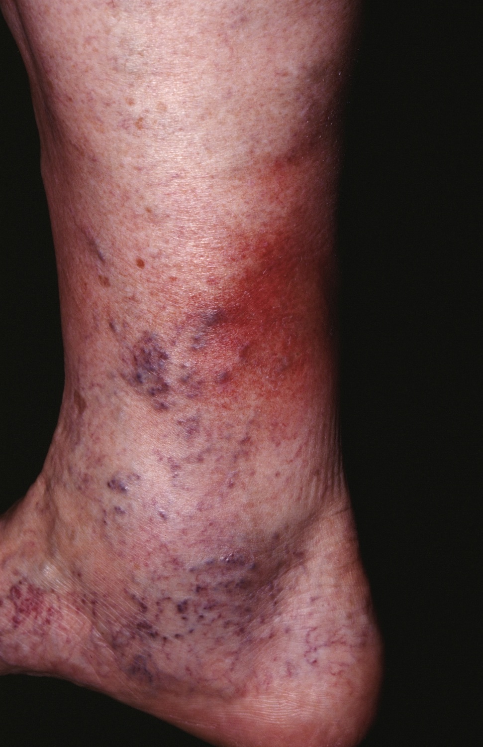
In the acute phase, a tender, inflamed, plaque occurs. Note the varicose veins/ankle flare indicating venous hypertension.
In the acute phase, a tender, inflamed, plaque occurs. Note the varicose veins/ankle flare indicating venous hypertension.
Lipodermatosclerosis is a thickening and inflammation of the skin about the medial lower leg. It results from chronic venous hypertension, usually in the setting of venous insufficiency.
One or both lower legs are affected. In the acute phase, a tender, inflamed, plaque occurs on the medial ankle. Over time, the skin becomes indurated and hardened. The demarcation between the edematous skin above and the indurated and bound-down skin below is relatively distinct. A drop-off is often noted. The shape of the leg is that of an inverted bottle. Areas of atrophy blanche are common. Varicose veins and an ankle flare are typical. A brownish color along with hemosiderin deposition is often seen. Pain is often a prominent feature. Ulceration may occur.
The diagnosis is usually a clinical one. One may consider a biopsy to verify the diagnosis, but healing will be slow and difficult. It should be avoided if possible. Occasionally, this condition is confused with Vilanova disease. Also, make sure the patient is not on pemetrexed, a cancer drug, which can cause a nearly identical condition (pemetrexed-induced inflammatory and sclerotic edema).
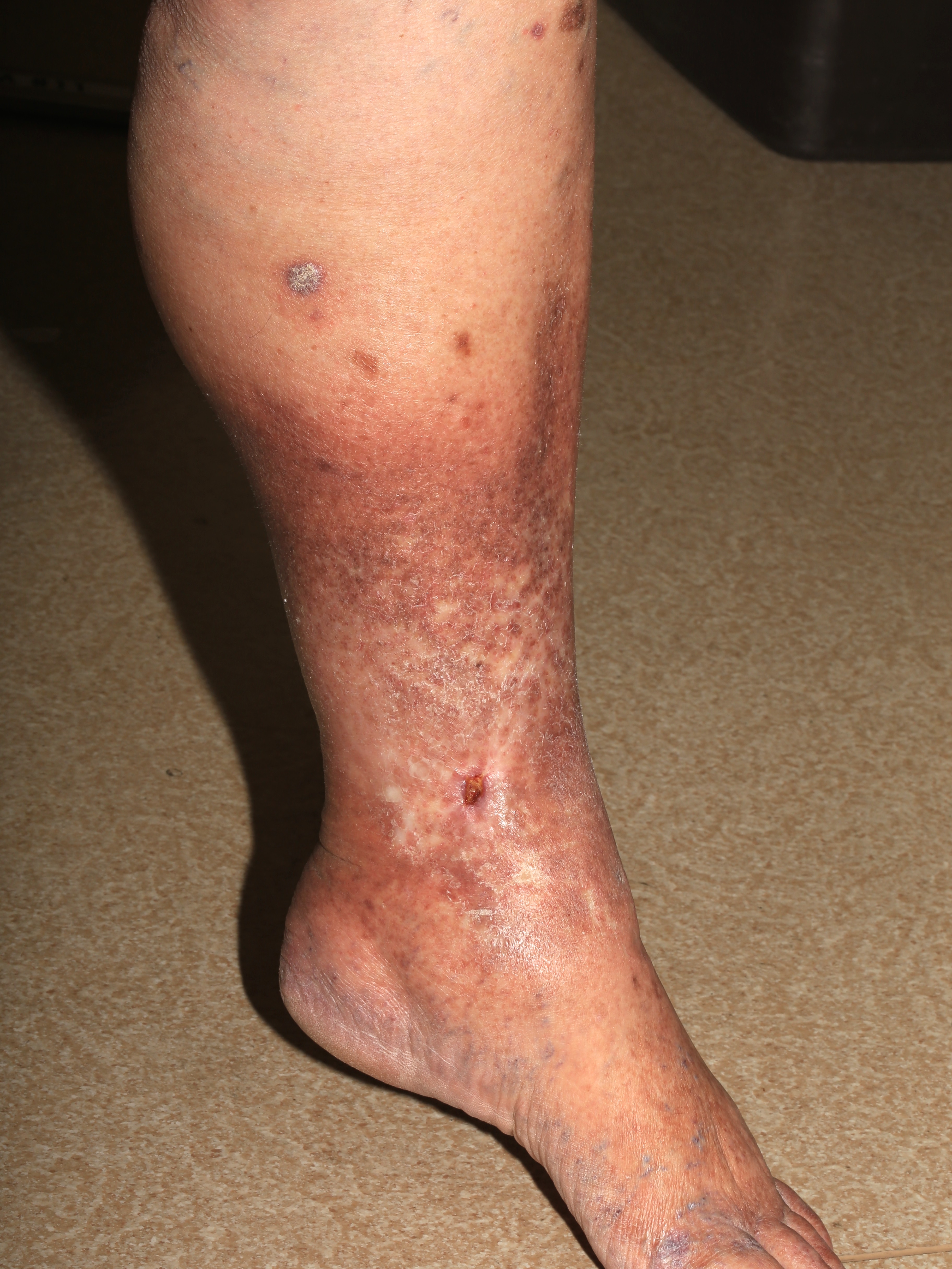
Circumferential induration, thickening and hyperpigmentation about the lower leg and ankle in a patient with more advanced lipodermatosclerosis.
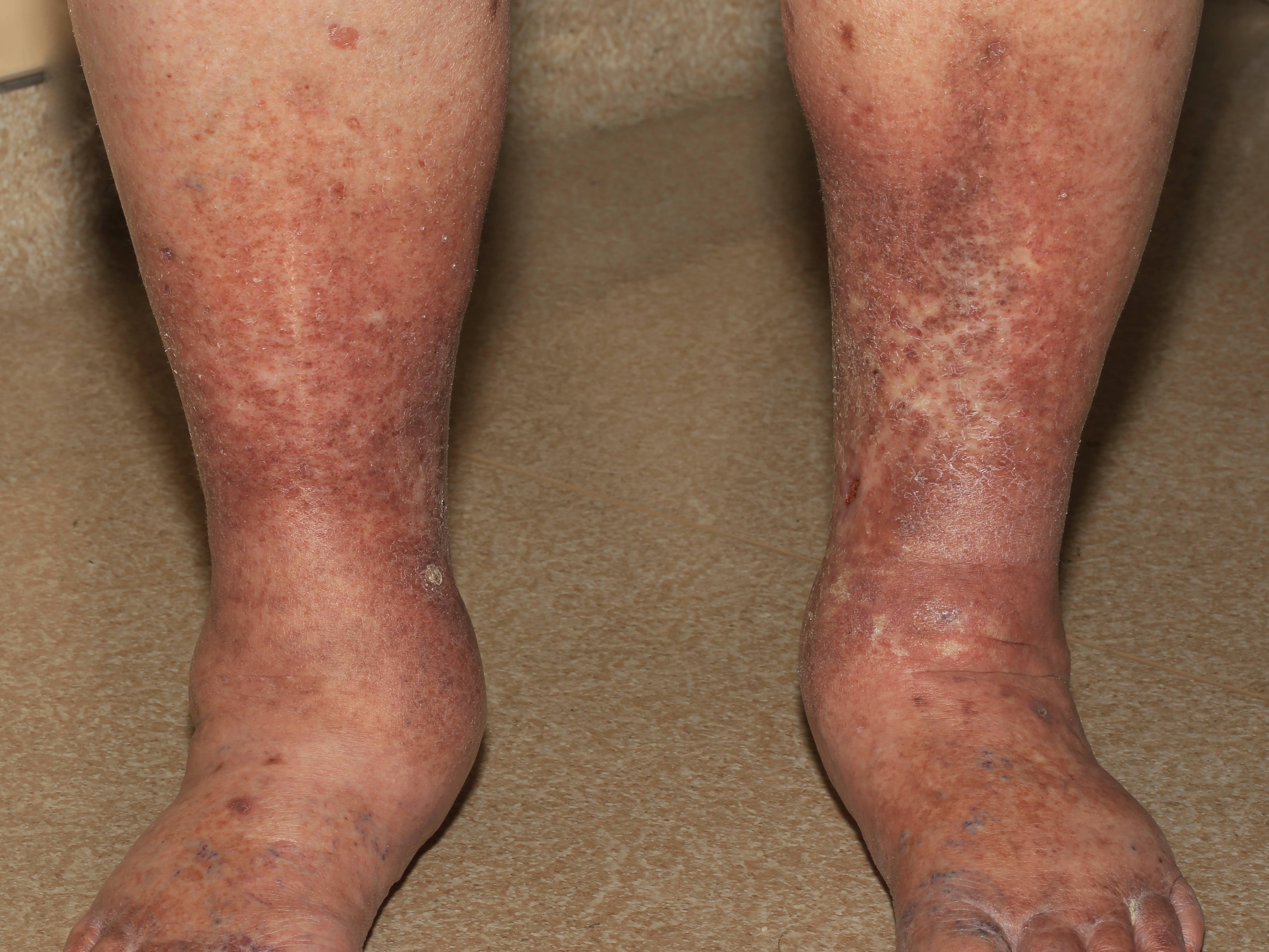
One or both legs may be affected.
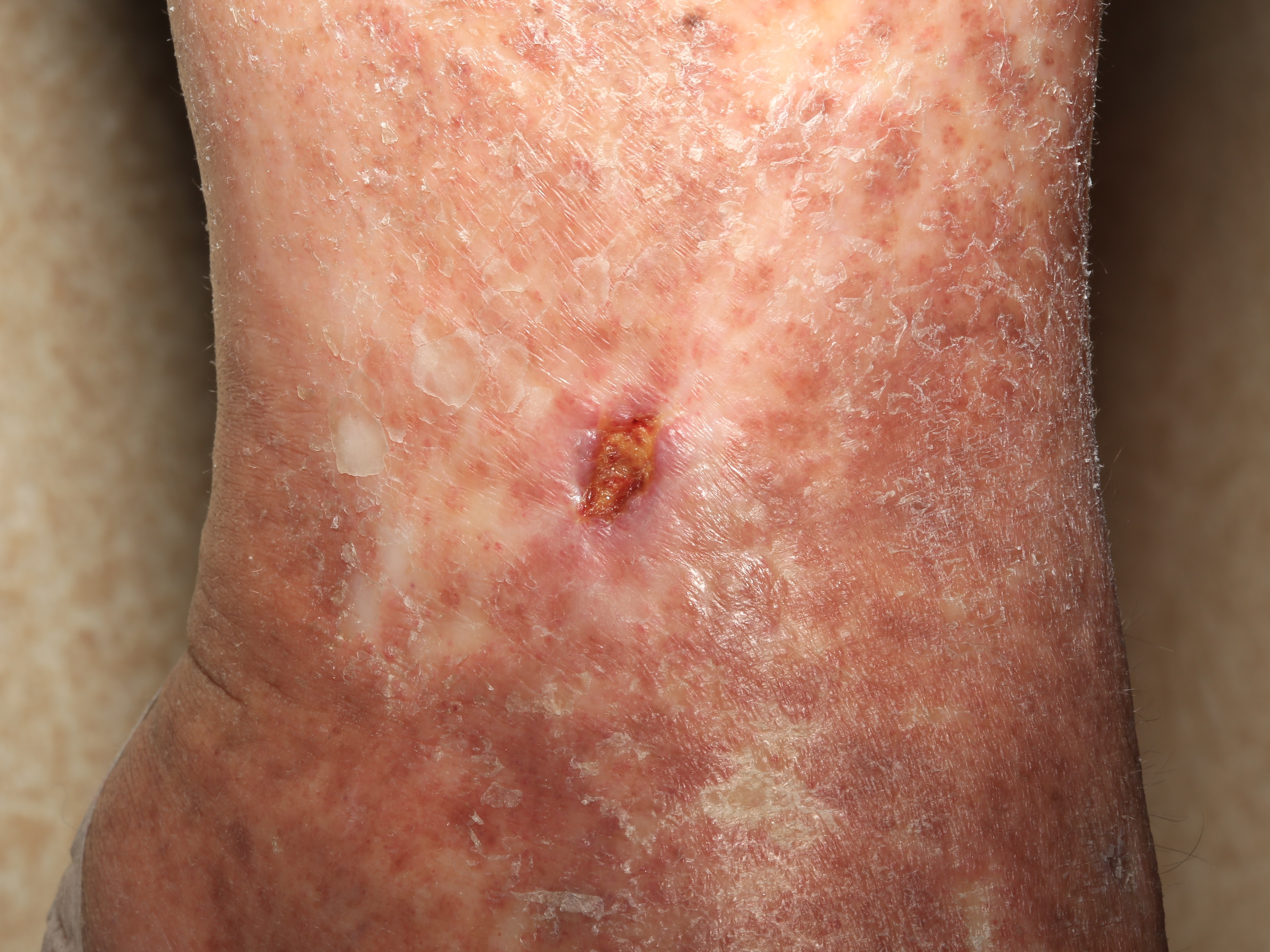
Skin breakdown may occur. Note the white stellate scars indication hypoxic damage.

Homepage | Who is Dr. White? | Privacy Policy | FAQs | Use of Images | Contact Dr. White