AMELANOTIC MELANOMA
The amelanotic melanoma (AMM) is a melanoma devoid of pigment. The correct diagnosis may not be suspected early on. This is why all lesions presumed to be pyogenic granulomas should be sent for histologic examination. Sometimes, an AMM may resemble a basal cell carcinoma. The AMM typically occurs in adulthood with a peak incidence in the fifth decade. They most commonly occur in the sun-exposed areas.
- Rapidly growing (6x faster than regular melanoma) but non-descript.
- Pink, "When you see pink, stop and think!"
- Sometimes has a hint of pigment.
- AMM represents a significant problem in terms of patient education and screening for melanoma as it does not fit the classic ABCDE criteria.
- Factors associated with increase risk of developing an AMM in one study included the absence of back nevi, presence of many freckles, a sun-sensitive phenotypic index and prior AMM.
Clinical
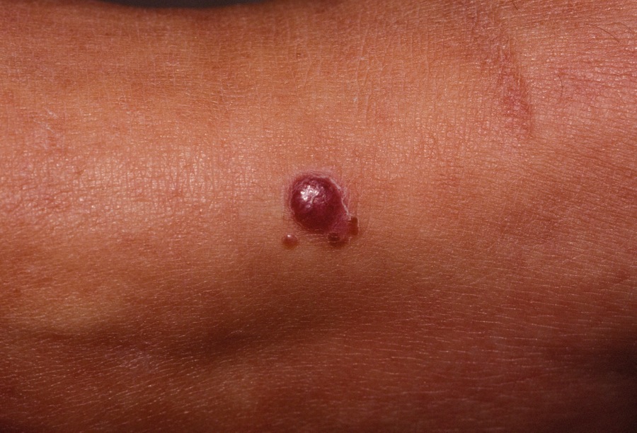
The lesion typically is a relatively rapidly growing pink, red or vascular-appearing papulonodule. ("When you see pink, stop and think"). Early lesions are often pink, symmetric and non-descript. Larger lesions often ulcerate or bleed. At times, there is a telltale bit of pigment at the base.
RegionalDerm
Homepage | FAQs | Use of Images | Contact Dr. White
It is not the intention of RegionalDerm.com to provide specific medical advice, diagnosis or treatment. RegionalDerm.com only intends to provide users with information regarding various medical conditions for educational purposes and will not provide specific medical advice. Information on RegionalDerm.com is not intended as a substitute for seeking medical treatment and you should always seek the advice of a qualified healthcare provider for diagnosis and for answers to your individual questions. Information contained on RegionalDerm.com should never cause you to disregard professional medical advice or delay seeking treatment. If you live in the United States and believe you are having a medical emergency call 911 immediately.
