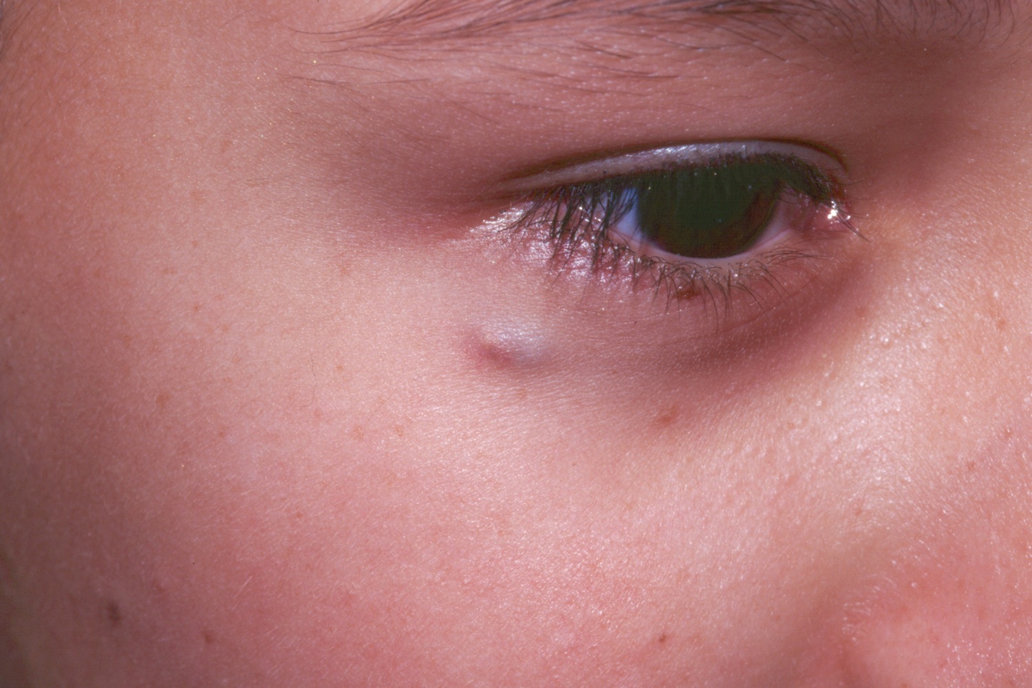
A 5-year-old girl with this rock hard dermal nodule. She was refered to a pediatric surgeon for excision.

A 5-year-old girl with this rock hard dermal nodule. She was refered to a pediatric surgeon for excision.
The pilomatricoma (PM) is a calcified tumor which seems to have its origin from the hair matrix cells. It may occur at any age, most commonly between 0 and 20 years of age, but there is another peak in the 50-70 age range.
A hard, irregular, dermal or subcutaneous tumor in a child, often on the head or neck, is characteristic. The typical lesion is between 1.0 and 1.5 cm in size. Females are more commonly affected. The overlying skin may be red, bullous, or flesh-colored. The tumor is often rock hard in consistency and an irregular surface may be appreciated. Usually the skin over the lesion is smooth and stretching the skin may show its multifaceted nature--the so called "tent sign." Occasionally, the skin may be keratotic, resembling an SCC or telangiectatic, resembling a BCC. Rarely, the overlying skin may wrinkle, a result of secondary anetoderma. Histologic studies suggest that the anetodermic pilomatricoma seems to arise from mechanical trauma to the overlying skin. If the skin is too thin, it may perforate allowing the extrusion of calcium.
A perfectly spherical nodule is more likely to be an epidermal inclusion cyst (EIC). The PM tends to be harder and with "bumpy" edges. Also, the PM is much more common in children whereas the EIC is more common in adults. Rapid growth should prompt a biopsy as rarely, pilomatrix carcinoma may occur.
Pilomatricomas may be multiple and familial or associated with myotonic dystrophy. Pilomatricoma-like changes have occurred in the cysts of Gardner syndrome.
Homepage | Who is Dr. White? | Privacy Policy | FAQs | Use of Images | Contact Dr. White